Radiomics-based machine learning models to distinguish between metastatic and healthy bone using lesion-center-based geometric regions of interest
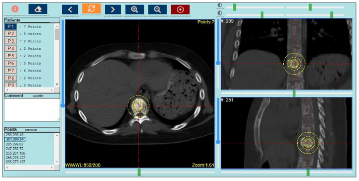 Image credit: Hossein Naseri
Abstract
Radiomics-based machine learning classifiers have shown potential for detecting bone metastases (BM) and for evaluating BM response to radiotherapy (RT). However, current radiomics models require large datasets of images with expert-segmented 3D regions of interest (ROIs). Full ROI segmentation is time consuming and oncologists often outline just RT treatment fields in clinical practice. This presents a challenge for real-world radiomics research. As such, a method that simplifies BM identification but does not compromise the power of radiomics is needed. The objective of this study was to investigate the feasibility of radiomics models for BM detection using lesion-center-based geometric ROIs. The planning-CT images of 170 patients with non-metastatic lung cancer and 189 patients with spinal BM were used. The point locations of 631 BM and 674 healthy bone (HB) regions were identified by experts. ROIs with various geometric shapes were centered and automatically delineated on the identified locations, and 107 radiomics features were extracted. Various feature selection methods and machine learning classifiers were evaluated. Our point-based radiomics pipeline was successful in differentiating BM from HB. Lesion-center-based segmentation approach greatly simplifies the process of preparing images for use in radiomics studies and avoids the bottleneck of full ROI segmentation.