The use of dose surface maps as a tool to investigate spatial dose delivery accuracy for the rectum during prostate radiotherapy
 Image credit: Haley Patrick
Abstract
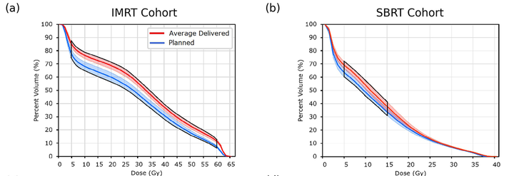
Purpose: This study aims to address the lack of spatial dose comparisons of planned and delivered rectal doses during prostate radiotherapy by using dose-surface maps (DSMs) to analyze dose delivery accuracy and comparing these results to those derived using DVHs. Methods: Two independent cohorts were used in this study: twenty patients treated with 36.25 Gy in five fractions (SBRT) and 20 treated with 60 Gy in 20 fractions (IMRT). Daily delivered rectum doses for each patient were retrospectively calculated using daily CBCT images. For each cohort, planned and average-delivered DVHs were generated and compared, as were planned and accumulated DSMs. Permutation testing was used to identify DVH metrics and DSM regions where significant dose differences occurred. Changes in rectal volume and position between planning and delivery were also evaluated to determine possible correlation to dosimetric changes. Results: For both cohorts, DVHs and DSMs reported conflicting findings on how planned and delivered rectum doses differed from each other. DVH analysis determined average-delivered DVHs were on average 7.1% ± 7.6% (p ≤ 0.002) and 5.0 ± 7.4% (p ≤ 0.021) higher than planned for the IMRT and SBRT cohorts, respectively. Meanwhile, DSM analysis found average delivered posterior rectal wall dose was 3.8 ± 0.6 Gy (p = 0.014) lower than planned in the IMRT cohort and no significant dose differences in the SBRT cohort. Observed dose differences were moderately correlated with anterior-posterior rectal wall motion, as well as PTV superior-inferior motion in the IMRT cohort. Evidence of both these relationships were discernable in DSMs. Conclusion: DSMs enabled spatial investigations of planned and delivered doses can uncover associations with interfraction motion that are otherwise masked in DVHs. Investigations of dose delivery accuracy in radiotherapy may benefit from using DSMs over DVHs for certain organs such as the rectum.